Lumbar Laminotomy and Foraminotomy
Learn what is involved in a minimally invasive lumbar laminotomy and foraminotomy performed on your lower back.
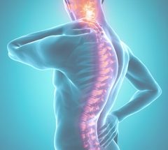
The spinal column consists of vertebrae separated by discs. Each vertebra consists of a main body (vertebral body) in the front and a bony canal (spinal canal) in the back. Between each of the bodies is a disc. The bony canal in the back contains the spinal cord and nerves. The roof of this canal is called the lamina. The spinous process is a bone between the laminae. This is the bone you feel when you press on your spine. The nerves exit the spinal canal through a small window called the foramina.
A laminectomy is a procedure when the whole lamina is removed. This is typically performed in an open (i.e., not minimally invasive) fashion to decompress the nerves. This procedure leaves the spinal canal open, and can be quite traumatic because the muscles, tendons, and ligaments that are normally attached to the spine must be stripped off and the entire lamina and spinous process are typically removed. The surgeons at Orthopedics International rarely have to perform open laminectomies, and believe it will be an outdated procedure in the near future.
A minimally invasive laminotomy, on the other hand, is a procedure that involves removal of a much smaller area of the lamina on one or both sides creating small holes that decompress the spinal cord and nerves. Unlike the laminectomy, a laminotomy can be performed in a minimally invasive fashion. Although the amount of bone that is removed is much less compared to an open laminectomy the surgical goals can still be achieved. Outcomes of a laminotomy from a functional and anatomic perspective are equivalent to the larger more open procedure of a laminectomy. The laminotomy is the procedure of choice for the minimally invasive spine surgeons at Orthopedics International.
A minimally invasive foraminotomy is a procedure that opens up the foramina (small window) where the nerves exit the spinal canal. When they become narrowed (stenotic), this procedure takes the pressure off of the exiting nerve root. You can think of this procedure as a functional “Roto-Rootering” of the window where the nerve leaves the spinal canal.
What conditions are treated with a laminotomy and foraminotomy?
Spinal stenosis (narrowing of the spinal canal or foramina) is the most common reason why patients undergo a laminotomy and/or foraminotomy. Over time, the spinal canal and/or foramina can become stenotic when the ligaments between the laminae thicken and the facet joints become arthrtic and overgrown. Disc bulges can also contribute to this narrowing. Often discrete bone spurs will form pinching a nerve. A person can also be born with a small spinal canal; this is called congenital spinal stenosis. Less common conditions that can lead to stenosis and require a decompression include facet cysts, fractures, and tumors.
What is the cause of spinal stenosis?
It is unclear why some people develop spinal stenosis and others do not. As previously mentioned, soft tissue and bony structures can narrow the areas where the neural elements live in the spinal canal. When the narrowing occurs in the spinal canal, we call this central stenosis. When narrowing occurs in the foramina, we call this foraminal stenosis. Spinal stenosis is a common problem in people over 50 years old. It most commonly occurs in the mid to lower lumbar spine and women are typically affected more often than men.
The pressure on these nerves can increase when you stand or walk, thereby causing an increase in your back pain and/or leg pain. The leg symptoms are quite varied, ranging from mild aching to severe fatigue. Leg pain, buttock pain, pins-and-needles sensations, and numbness are also common symptoms in patients with spinal stenosis. Your ability to walk may also be limited to a few blocks.
The goal of laminotomies and foraminotomies is to open up the areas for your nerves by removing the excess bone and soft tissue.
What is involved with the surgery?
A one-inch incision is made in your back at the operative level. A needle is then inserted through this skin incision onto your spine in the area surgery is to be performed. A series of dilators are placed so as to spread the muscle tissue rather than cut it.
Once the largest dilator has been placed, a one-inch in diameter tube-shaped retractor is inserted over the last dilator. The needle and dilators are then removed and the tube retractor is attached to a clamp to secure it in place. The surgery is then performed through this tube. A microscope is used for better visualization and safety.
As little bone is removed as possible to prevent instability of the spinal column. The surgery takes approximately an hour per level depending on the severity of the stenosis. Most patients are able to go home the same day as surgery.
What symptoms are relieved with a laminotomy and foraminotomy?
Symptoms in the buttocks and legs are most reliably relieved with surgery. These symptoms include pain, numbness, tingling, aching, heaviness, weakness, and inability to walk.
What is the overall success rate?
The success rate for this procedure is around 80% to 90%. The success of the procedure, however, often depends on your specific pathology. One of the first things you should notice a few weeks after the procedure is an improvement in your leg pain and your ability to walk. If you had leg weakness before the surgery, you will be enrolled in a physical therapy program to help strengthen your legs after your procedure. Resolution of numbness, however, tends to be somewhat unpredictable.
What can I do when I go home?
You can be as active as your pain allows. You can ride in a car, climb stairs, and generally perform your normal activities for the first month after surgery. At the one-month mark, you may be enrolled in a structured physical therapy program to include core strengthening and neutral spine exercises. You will also be allowed to begin low impact aerobic activities (e.g., elliptical training, recumbent bicycling, swimming, etc.) at this time point. Three months after surgery, you can begin higher impact aerobic activities such as running, jumping, and non-contact sports. Six months after surgery, you will be free to participate in any physical activity (including contact sports and activities) without restriction.
Should I have the surgery?
If the symptoms in your legs are simply annoying and mild you probably should not undergo the operation. However, if your quality of life is significantly affected by the symptoms associated with spinal stenosis, this procedure can often be life changing. Since the procedure is considered elective in the majority of cases, however, it is your responsibility to determine if your symptoms warrant the risk of surgery.
Feel free to discuss any further questions or concerns with your physician.


